SOCIAL MEDIA
Portuguese Medical Association's Scientific Journal

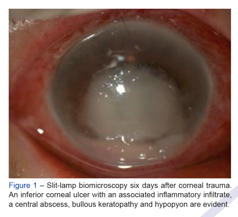
Introduction: Infectious keratitis is an important cause of visual loss. The purpose of this study was to investigate anterior segment optical coherence tomography patterns in infectious keratitis and evaluate the role of this tool in the early management of this disorder.
Material and Methods: In this cross-sectional study, we included patients with proven infectious keratitis, either by culture or therapeutic trial. Subjects underwent baseline anterior segment optical coherence tomography (Spectralis® anterior segment module, Heidelberg Engineering, Germany) performed by the same operator. We used anterior segment optical coherence tomography vertical and horizontal raster default scans with 6.0 mm scan lines.
Results: Twenty-five patients (14 men and 11 women) were included. The most common risk factors identified were ocular trauma (11 cases) and contact lens wear (7 cases). Fifteen patients presented bacterial infection; three, fungal infection; two parasitic infection; and five cases presented a negative microbiological culture. Anterior segment optical coherence tomography depicted nine distinct morphological patterns.
Discussion: Anterior segment optical coherence tomography allows the depth of corneal involvement to be assessed. When the only patterns identified were hyperreflective stromal lesion and stromal edema, the visual outcome was better. Cystic spaces were present
in severe bacterial keratitis.
Conclusion: Anterior segment optical coherence tomography can complement biomicroscopy, allowing for a better characterization of corneal involvement at presentation that can help in staging and providing useful prognostic information.