SOCIAL MEDIA
Portuguese Medical Association's Scientific Journal

A 78 year-old man, being studied for wasting syndrome with unspecific symptoms, performed a contrast-enhanced computed tomography (CT) scan, revealing stomach hipodensity and distension with linear hipodensity in the fundus wall, promptly visible on the scout view (Fig. 1A). On the axial and multiplanar reconstruction images there are findings of gastric pneumatosis (intramural gas) (Fig.s 1B, 1C, 1D), a rare finding, the stomach being the least common location of intramural pneumatosis in the gastrointestinal tract. Several causes have been described, emphysematous gastritis being the most lethal.
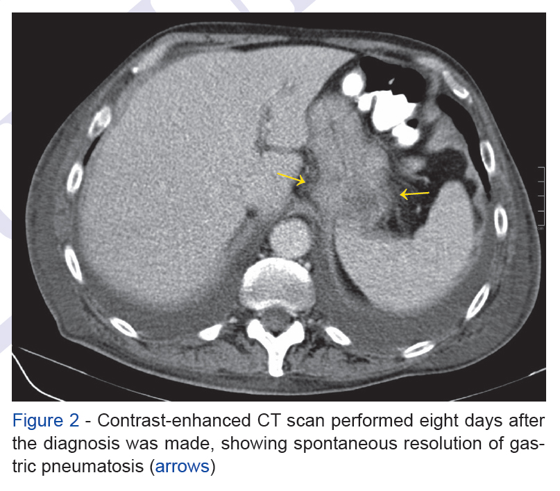
There were no relevant clinical or analytical findings, no signs of pneumatosis in other segments of the gastrointestinal tract or the portal vein. The patient underwent an upper gastrointestinal endoscopy two days before, where two biopsies were performed in the duodenal bulb. This prompted the diagnosis of endoscopy-induced gastric pneumatosis. Therapy was supportive and the control CT performed eight days later revealed spontaneous resolution (Fig. 2).
Read the whole article here (english only)