SOCIAL MEDIA
Portuguese Medical Association's Scientific Journal

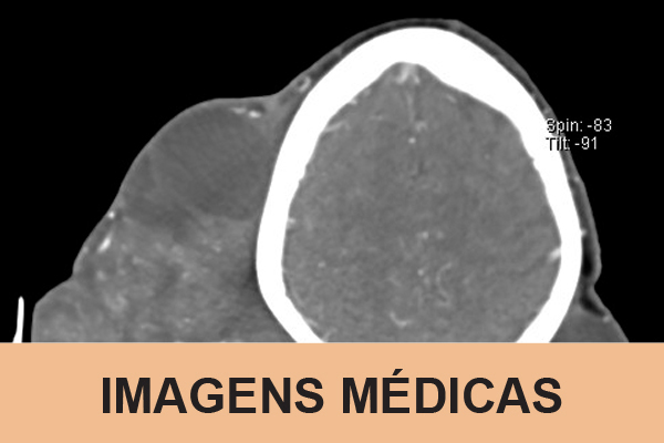
A 76-year-old female presented with a massive, exophytic, multinodular and well-circumscribed lesion of the scalp, with two years of evolution, measuring 28.3 x 25.4 x 22.9 cm.
The lesion was partially necrotic with both solid and cystic areas (Fig. 1). A computed tomography scan did not reveal distant dissemination, nor bone invasion of the cranial vault (Fig. 2). She underwent extended excision of the lesion with preservation of the periosteum and reconstruction of the defect with a partial-thickness skin graft. The histological examination revealed a benign proliferative trichilemmal tumor (PTT) without atypia.